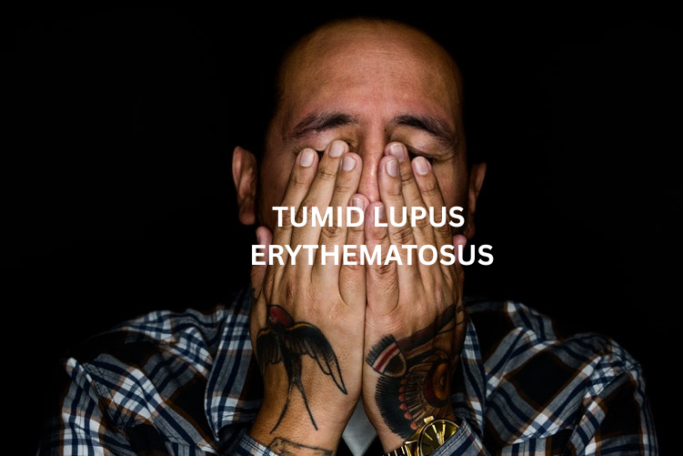
Tumid lupus erythematosus (TLE) is a rare photosensitive sub type of cutaneous lupus erythematosus (CLE). It causes a person to develop red or violet smooth raised lesions on their skin that can resemble urticaria (hives).
A key feature differentiating it from discoid lupus erythematosus (DLE) is the absence of epidermal changes like atrophy, scaling or scarring after the lesions resolve. Lesions frequently appear on sun-exposed skin, such as the face, neck, upper chest (‘v area’) and arms.
TLE is rarely associated with Systemic Lupus Erythematosus (SLE), with a weak link to serological abnormalities.
Diagnosis

A biopsy of a lesion is crucial for diagnosis, and shows characteristic changes including lymphocytic infiltration around blood vessels and skin appendages and abundant mucin (a substance that provides the "swollen" appearance) in the dermis.
A dermatologist will also examine the skin for the characteristic non-scarring plaques in photosensitive areas. The absence of epidermal changes like scaling, atrophy, or follicular plugging helps differentiate TLE from other forms of cutaneous lupus.
Blood tests for systemic lupus, such as the antinuclear antibody (ANA) test, are typically negative or show low titres in TLE patients.
Causes
Genetic predisposition is thought to play a role, as a family history of lupus or autoimmune disease can increase risk.
Sunlight (specifically UVA and UVB) is a major trigger that can induce lesions.
Other environmental triggers such as smoking and certain medications (e.g., TNF-alpha inhibitors, thiazide diuretics), have been linked to TLE.
Treatment
Sun Protection: Sun exposure is a major trigger for the condition and can cause flare-ups or worsen existing lesions. Sunscreen with SPF 30+, protective clothing and avoiding peak sunlight hours are essential.
Medications: Antimalarial drugs, such as hydroxychloroquine, are often effective. Topical corticosteroids may also be used.
Stopping smoking is strongly advised, as it can worsen lupus symptoms and reduces the effectiveness of antimalarial drugs.
Prognosis
TLE generally has a favourable prognosis, with lesions often responding well to treatment and regressing without scarring. TLE lesions can also resolve completely with treatment or sometimes spontaneously. The risk of progression to systemic lupus erythematosus (SLE) is significantly lower than with other forms of lupus.
Further Information
For further research (National Library of Medicine) click here.
For further research (Teen with TLE) click here.
Photograph courtesy of DermNet.
